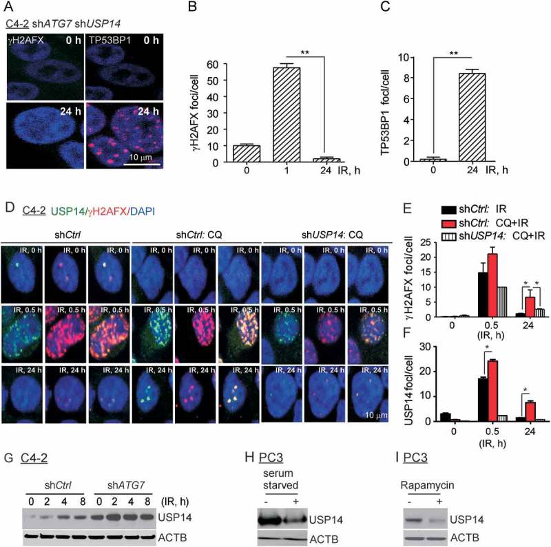
Figure 2.

USP14 disrupts DDR signaling in autophagy-deficient cells. (a–c) Confocal immunostaining and graphical representation of γH2AFX and TP53BP1 foci following IR treatment in C4-2 cells co-expressing shATG7 and shUSP14. Nuclei were stained with DAPI. (d–f) Confocal immunostaining and graphical representation of γH2AFX and USP14 foci following IR+/− CQ treatment in C4-2 cells expressing shCtrl and shUSP14. Nuclei were stained with DAPI. Data shown are the means ± SEM (n = 2); P < 0.05 *, P < 0.01 **. (g) Western blot analysis for USP14 in shCtrl-expressing vs shATG7-expressing C4-2 cells following IR treatment for the indicated time. ACTB/β-actin was used as a loading control. Western blot analysis for USP14 in PC3 cells treated with (h) serum starvation and (i) rapamycin. ACTB/β-actin was used as a loading control.
