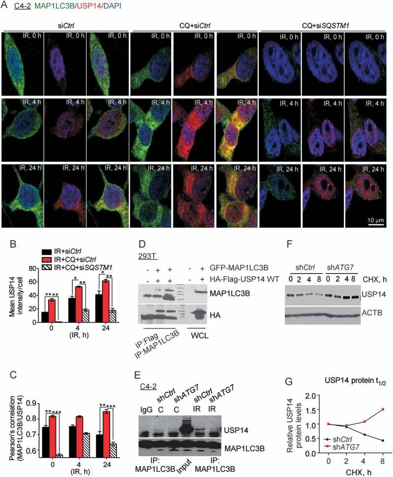
Figure 4.

USP14 is a substrate of autophagy. (a–c) Representative confocal images and quantification of MAP1LC3B and USP14 levels and colocalization in C4-2 cells treated as indicated. Nuclei were stained with DAPI. (d) 293T cells were transfected with the indicated constructs, followed by immunoprecipitation as indicated and immunoblotting using the anti-MAP1LC3B and -HA antibodies. The corresponding WCLs were used as input controls and probed with the indicated antibodies. (e) MAP1LC3B was immunoprecipitated from C4-2 cells stably expressing shCtrl or shATG7, treated as indicated, followed by western blotting using anti-USP14 and -MAP1LC3B antibodies. (f) Western blot analysis and (g) relative protein quantification for USP14 in shCtrl vs shATG7-expressing C4-2 cells following cyclohexamide (CHX) treatment for the indicated time. ACTB/β-actin was used as a loading control. Data shown are the means ± SEM (n = 2); P < 0.05 *, P < 0.01 **, P < 0.001***.
