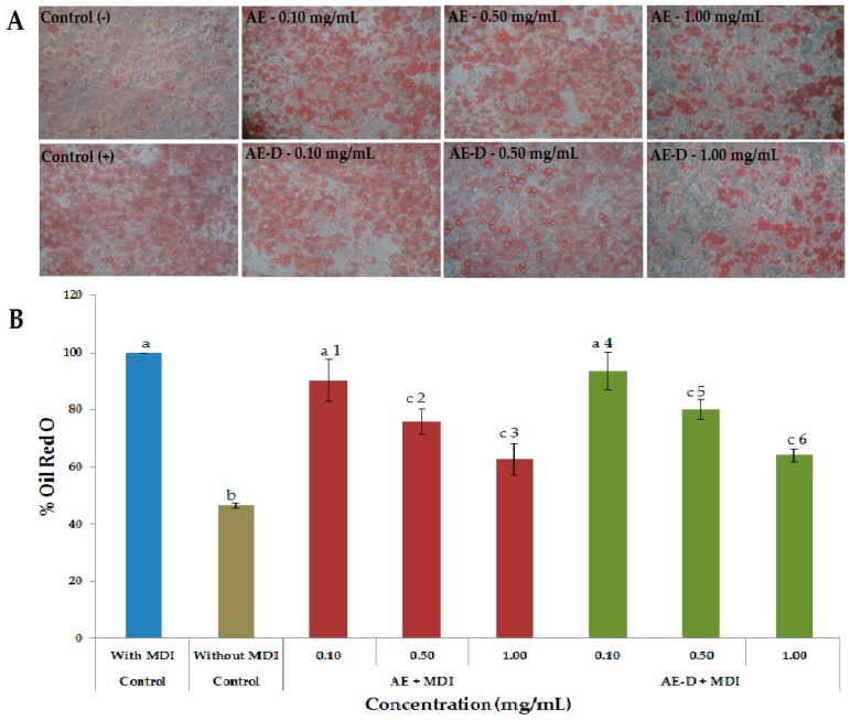
Figure 3.
Adipocytes stained with the dye Oil Red O after 15 days after the onset of differentiation. (A) Intracellular lipid accumulation in 3T3-L1 cells treated with adipogenic medium (MDI) and different concentrations of AE or AE-D. After 15 days of treatment the cells were stained with oil Red O, observed with a microscope and photographed (20× magnification). (B) Baccharis trimera extracts inhibited the differentiation of 3T3-L1 preadipocytes into mature adipocytes. Oil Red O dye was used to stain the adipocytes 15 days after treatment with the extracts. Stained oil droplets were dissolved with isopropanol and evaluated by spectrophotometric analysis at 510 nm. The optical density in cells treated only with MDI was taken as 100% of relative lipid content. Values are expressed as mean ± standard deviation (n = 3). Letters a,b,c indicate significant differences between different concentrations with the control. Numbers 1,2,3,4,5,6 indicate significantly differences between different concentrations of same extract according to one-way analysis of variance (ANOVA) followed by the Student’s t-test (p < 0.05).

