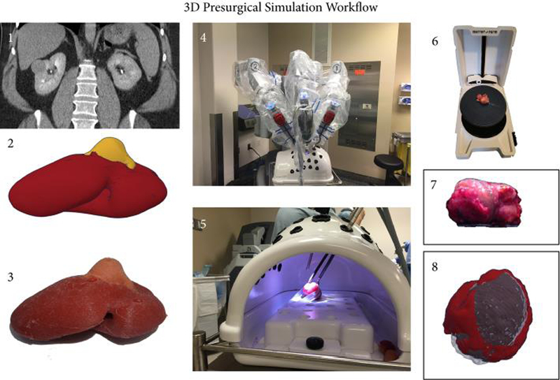
Figure 1.

Workflow for the Presurgical Simulation. A patient specific 3D digital model is reconstructed on the basis of the available preoperative axial imaging (CT or MRI) (1-2) and then converted into a cast silicone soft tissue model as described below (3). Robotic-assisted laparoscopic enucleation of the kidney tumor is rehearsed for all patients using the 3D printed model in a three-arm configuration (4-5). Both the resected model tumor and the patient’s actual resected tumor are 3D laser scanned in the operating room for volumetric measurement (6-7). All scans are then reprocessed and each pair (specimen scan from 3D kidney model and patient) is digitally overlaid to visualize the concordance in mass contour.
