Figure 1:
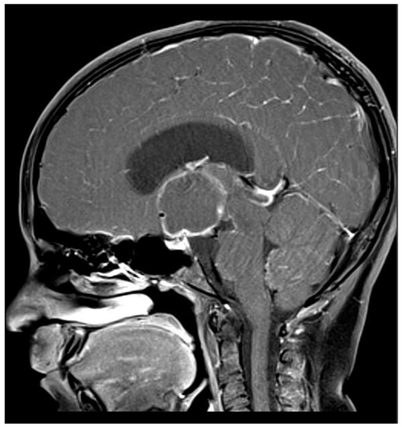
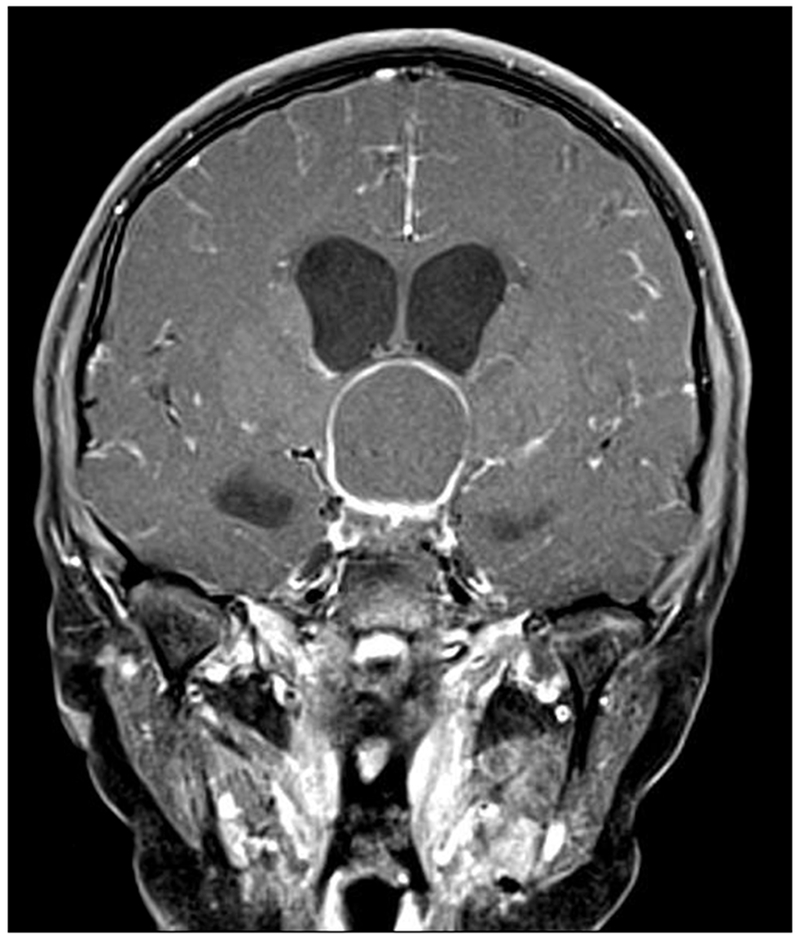
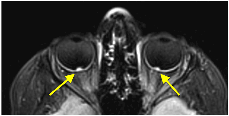
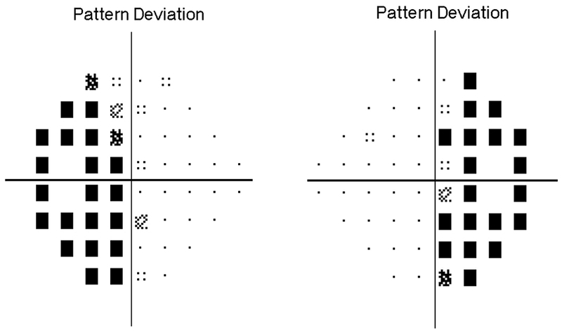
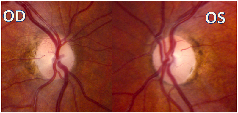
A 10-year-old girl presented with headaches, diplopia, nausea, and vomiting. Sagittal T1-weighted post-gadolinium with fat suppression magnetic resonance image (A) and coronal T1-weighted post-gadolinium with fat suppression image (B) revealed a large solid and cystic suprasellar mass resulting in obstructive hydrocephalus and compression of the optic chiasm. Ophthalmologic examination revealed bilateral sixth nerve palsies and moderate papilledema, which can be seen on the axial FLAIR post-contrast image (C, arrows). The patient underwent resection, and pathology returned consistent with craniopharyngioma. Compression of the optic chiasm led to a bitemporal hemianopia (D). Papilledema and compression of the optic chiasm led to eventual optic nerve pallor with peripapillary changes (E) after resolution of the disc edema.
