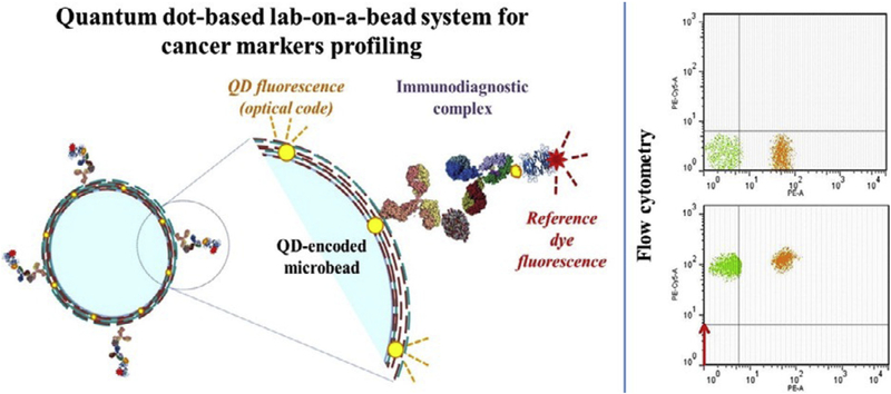
Fig. 10. QD-encoded microbeads for multiplexed cancer diagnosis.
(a) Schematic of the designed lab-on-a-bead system for detection of prostate-specific antigen (PSA).
(b) Flow cytometry dot plots of the suspension array based on two microbead populations carrying different fluorescent nanocrystal codes for simultaneous detection of free PSA and total PSA in clinical serum samples. A green (PE-A-negative, FITC-A-positive) microbead population was used for free PSA detection, and an orange (PE-A-positive, FITC-A-negative) microbead population was used for the total PSA detection Panel A Detection of two PSA forms in the serum of a healthy female donor (the PSA-negative control). Panel B. Detection of two PSA forms in the serum of a prostate cancer-positive male patient (the PSA-positive control). Red fluorescence shifts of different intensities in the PE-Cy5-A channel indicate binding of different amounts of PSA. Reprinted from [95] With permission. (For interpretation of the references to color in this figure legend, the reader is referred to the Web version of this article.)

