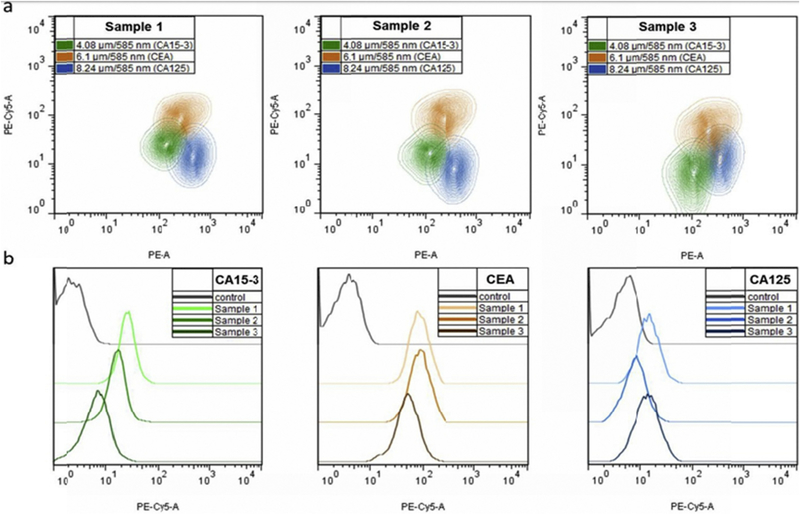
Fig. 9. Flow cytometry analysis of multiplexed QDebead microarrays used for simultaneous detection of CA 15e3, CE A, and CA 125, markers of female reproductive system tumors, in clinical serum samples.
(a) Simultaneous detection of three cancer markers in three individual serum samples from patients with different stages of breast cancer.
(b) Comparative histograms indicating different levels of each target marker in three analyzed serum samples in comparison with the control sample. FSC-A, bead size; PE-A bead optical code (QD 585 nm fluorescence); PE Cy5-A, the amount of the cancer marker detected (fluorescence of the streptavidin-Tri-COLOR visualization label). Reprinted from [93] With permission. (For interpretation of the references to color in this figure legend, the reader is referred to the Web version of this article.)

