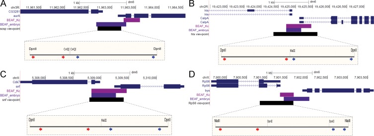
Fig 1. Locations of the 4C viewpoints.
(A) Chr3R scs’ viewpoint at the divergent CG3281 and aurA genes. (B) Chr2R hts viewpoint at the divergent hts and CalpA genes. (C) ChrX snf viewpoint at the divergent Cdk7 and snf genes. (D) ChrX RpS6 viewpoint at the divergent RpS6 and bys genes. The UCSC Genome Browser snapshots show the gene models (blue), BEAF peak limits from Kc cell (magenta) or embryo (purple) mapping, and the viewpoint region (black). The expanded views show the sites for the two restriction enzymes used, with the positions of inverse-PCR primers for the left and right sides of the viewpoints indicated by red and blue arrow pairs respectively.

