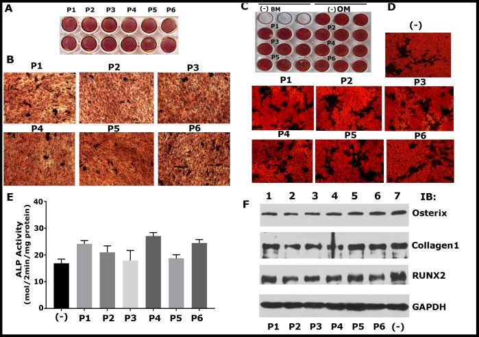
Fig 6. Mineralization and expression of osteogenic markers (Collagen, RUNX2, and Osterix) by osteoblasts are unaffected by sNT-LPL peptides (P1-P6).
(A-D) Mineralization was assessed by Alizarin Red Staining (ARS) in cells fixed at day 7 of culture. ARS of MC3T-E1 (A and B) and UMR-106 (C and D) cells cultured for seven days is shown. Each peptide treatment was done in duplicates for MC3T3 (A) and triplicates for UMR-106 (C) cells. The plates were scanned in an EPSON Perfection V200 Photo scanner (A and C). Representative magnified images of mineralized nodules are shown (B and D). Magnified images were taken in a phase contrast microscopy with a 10X and 20X objective for B and D, respectively. Cells were grown in osteogenic medium (OM), and basal medium (BM) without peptides (-) were used as controls. (E) Analysis of ALP activity in UMR 106 cells. Data shown are mean ± SD (n = 3). Minus (-) in C indicates cells grew in OM but untreated with the peptide. There is no significant statistical difference between the groups. (F) Western blot analysis for the expression of osteogenic biomarkers such as Collagen 1, RUNX2 and osterix. Lysates made from osteoblasts treated with indicated peptides for seven days were used for the analysis. Immunoblotting with a GAPDH antibody was used as loading control. Results in A-D and F represent one of the two experiments performed with the similar results.

