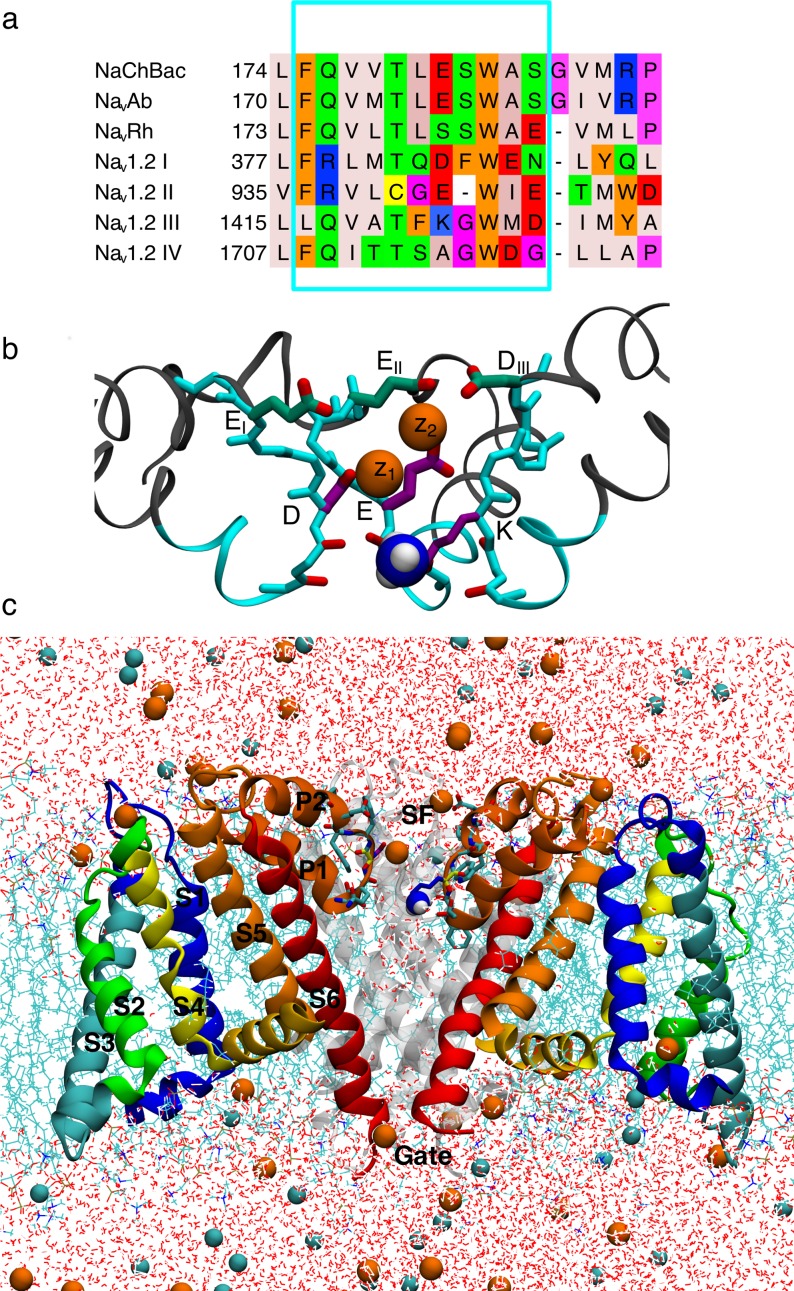
Fig 1. Human Nav1.2 model ion channel system.
a) Jalview [81] sequence alignment of bacterial NaChBac, NavAb and NavRh, with the four domains of human Nav1.2 (labeled DI-DIV). Amino acids are colored according to their properties using the Zappo coloring scheme [81]. Cyan box selection marks the grafted Nav1.2 residues. Red box selection marks the EEEE/DEKA signature sequences responsible for selectivity. b) SF and vestibule of the NavRh/Nav1.2 model with grafted residues in cyan. The residues in the inner DEKA and outer EEDD rings are indicated in purple and green, respectively, and the charged ammonium group of the Lys is shown as blue and white balls. c) NavRh/Nav1.2 channel embedded in a hydrated DPPC bilayer (cyan sticks), surrounded by water (red and white sticks) with Na+ and Cl- ions (orange and cyan balls). Three out of four monomers are shown (front subunit removed for clarity; rear subunit in gray), with VSD in green/blue/yellow and the PD in orange/red ribbons.

