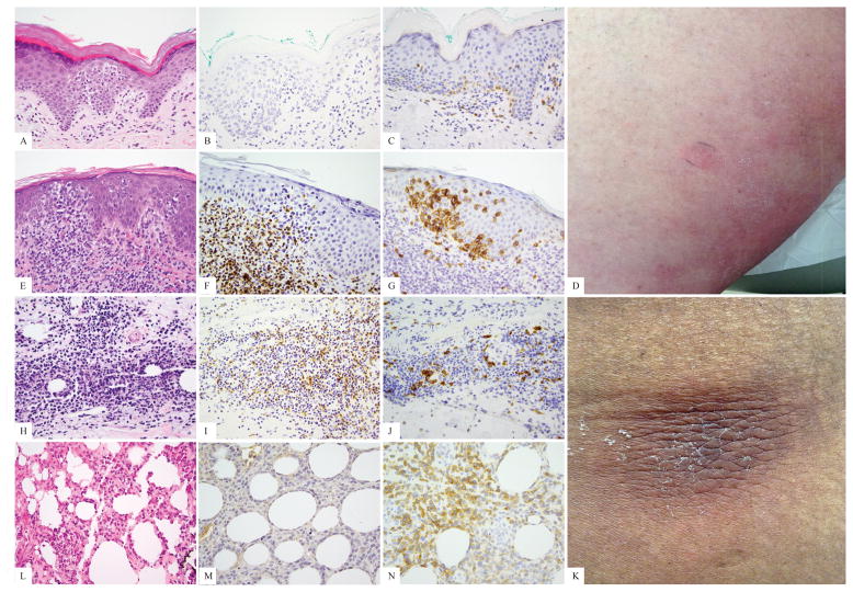
Figure 3.
Examples of primary cutaneous T-cell lymphomas with clinical behavior of mycosis fungoides or subcutaneous panniculitis like T-cell lymphoma, and prominent TCRγδ phenotype as shown by TCRδ immunohistochemistry. A–D, hematoxylin-eosin, TCRβ, TCRδ, and clinical patch lesions in a patient with a mycosis fungoides presentation and 84 months of follow-up; E–G, hematoxylin-eosin, TCRβ, and TCRδ in a recently diagnosed patient with a mycosis fungoides-like presentation; H–K hematoxylin-eosin, TCRβ, TCRδ, and clinical subcutaneous nodule in a panniculitic patient with indolent course through limited (several month) follow up; L–N, hematoxylin-eosin, TCRβ, and TCRδ in a panniculitic presentation with indolent course through 84 months.

