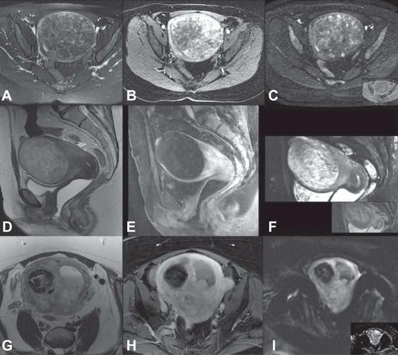
Figure 3.
Use of T2-weighted images, dynamic magnetic resonance imaging with contrast enhancement, and diffusion weighted imaging (with inset apparent diffusion coefficient map) for leiomyoma or leiomyosarcoma diagnosis. Benign leiomyoma: mildly heterogeneous low T2 signal (A), early and heterogeneous avid enhancement (B) no significant restricted diffusion (C). Degenerated leiomyoma: heterogeneous high T2-weighted signal (D) T1-weighted imaging with fat saturation shows homogeneous hypoenhancement (E) heterogeneous diffusion restriction (F). Leiomyosarcoma: infiltrative, ill-defined mass with intermediate T2 signal (G) enhancement of the viable tumoral tissue (H) and hyperintense on high B-value imaging with subsequent restricted dark signal on the apparent diffusion coefficient map (I).

