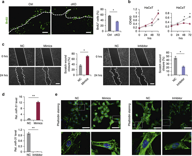
Figure 3. MiR-31 promotes proliferation and migration of keratinocytes.

(a) Immunostaining for BrdU in the wound edge keratinocytes in miR-31 cKO and control wounds on PWD 6 following 90 min BrdU pulse. Scale bar: 25 μm. (b) WST-8 assay showing human HaCaT keratinocyte growth changes in response to miR-31 mimics (blue) and inhibitor (red). (c) In vitro scratch assay showing changes in migration potential of HaCaT cells in response to miR-31 mimics and inhibitor. (d) qRT-PCR for miR-31 to test the transfection efficiency for miR-31 mimics and inhibitor. **p < 0.01. (e) Phalloidin staining (green) in HaCaT keratinocytes transfected with miR-31 mimics (left) and inhibitor (right). Scale bar: 25 μm. n =3 biological replicates for a-e.
