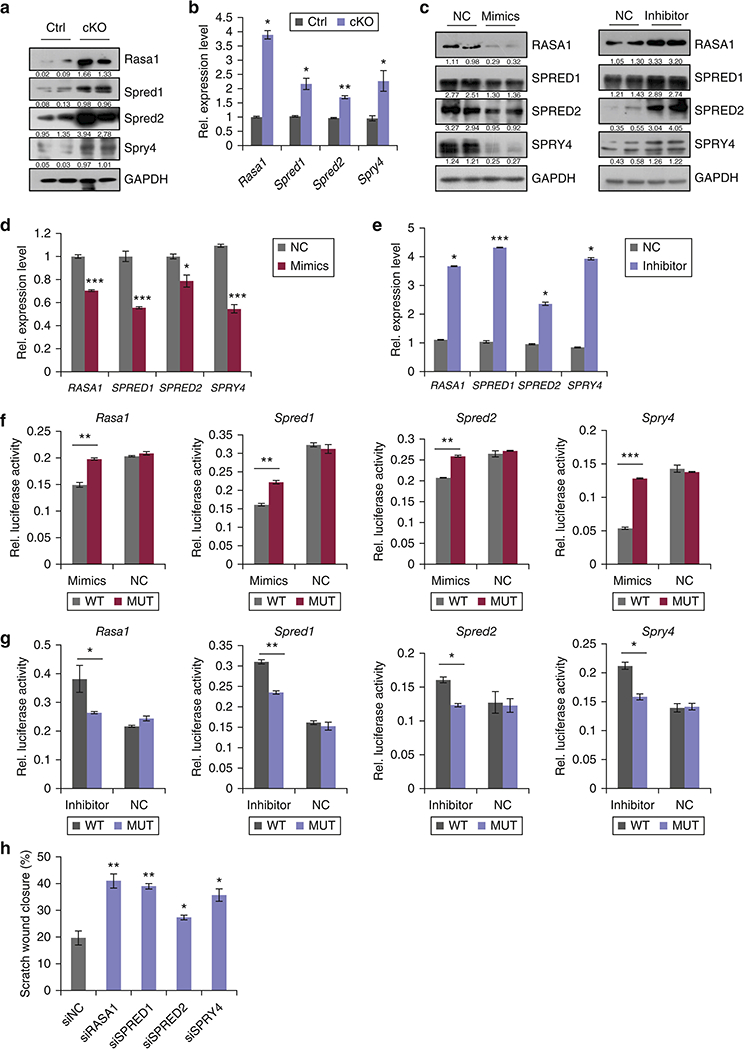
Figure 5. Identification of miR-31 direct targets.

(a,b) Western blot (a) and qRT-PCR (b) for Rasa1, Spred1, Spred2 and Spry4 in miR-31 cKO and control wounds on PWD 6. (c-e) Western blot for RASA1, SPRED1, SPRED2 and SPRY4 (c), and qRT-PCR for Rasa1, Spred1, Spred2 and SPRY4 (d,e) in HaCaT cells transfected with miR-31 mimics or inhibitor. (f,g) Luciferase reporter activity of WT and mutant 3’UTR constructs of Rasa1, Spred1, Spred2 and Spry4 in HaCaT cells transfected with miR-31 mimics (f) and miR-31 inhibitor (g). (h) Statistical analysis on wound closure in in vitro scratch assay shown in supplementary Figure S6c. n =3 biological replicates for a-h. *p< 0.05;**p<0.01;***p<0.001.
