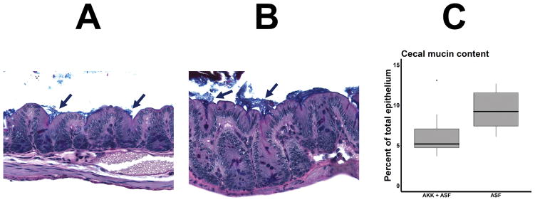
Figure 7.
Mucin thickness of transplanted mice. Cecal sections were stained with the Alcian Blue pH 2.5 + Periodic acid–Schiff (PAS) stain, and the area of mucus and epithelium were determined by manual tracing using Image Pro Plus v7.0. 7A and 7B depict representative sections of mice receiving ASF with (7A) or without (7B) supplemental A. muciniphila. Mucin stain appears as a blue layer (arrows) overlying the epithelium. A Boxplot summarizing the results is shown in 7C (n = 10 for A. muciniphila + ASF, n = 9 for ASF alone.)

