Figure 3.
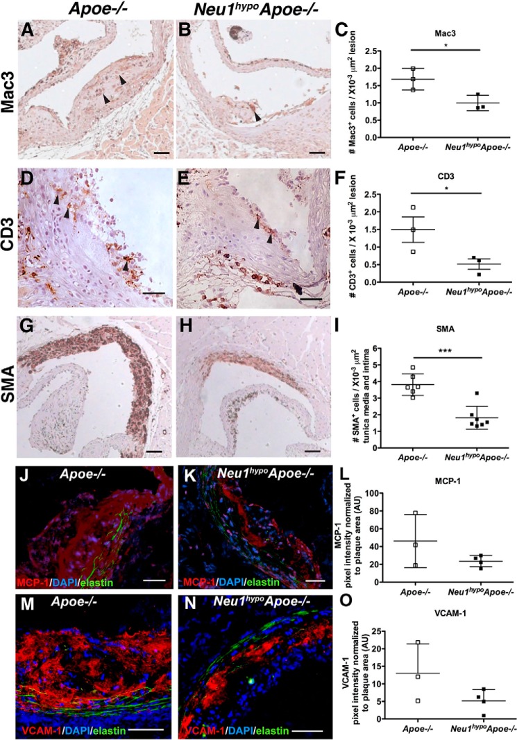
Assessment of cell types in the aortic sinuses of Apoe−/− and Neu1hypoApoe−/− mice. The infiltration of macrophages, T cells, and smooth muscle cells into the aortic sinus of 7-month-old Apoe−/− and Neu1hypoApoe−/− male mice was analyzed. Mac3 (A–C), CD3 (D–F), and SMA (G–I) were used to detect macrophages, T cells, and smooth muscle cells, respectively. Hematoxylin (blue) was used for nuclear staining, and representative sections are shown from male Apoe−/− and Neu1hypoApoe−/− mice. Arrowheads indicate immunoreactive Mac3+ and CD3+ cells. Scale bars = 50 μm. Neu1hypoApoe−/− male displayed a 41% reduction of Mac3+ cells (C) and a 66% reduction of CD3+ cells (F) in atherosclerotic lesions compared with controls. I, there was also a significant reduction of SMA+ cells in Neu1hypoApoe−/− mice compared with Apoe−/− mice. Individual data are presented with mean ± S.D. *, p < 0.05; ***, p < 0.001. J–L, monocyte chemoattractant protein (MCP-1) was detected in aortic sinus lesions of Apoe−/− (J) and Neu1hypoApoe−/− (K) mice using immunofluorescence. Scale bars = 50 μm. L, MCP-1 expression, expressed in arbitrary units (AU) was quantified by normalizing MCP-1 pixel fluorescence intensity to lesion area. Sialidase deficiency did not significantly alter MCP-1 expression. M–O, vascular cell adhesion protein 1 (VCAM-1) was detected in aortic sinus lesions of Apoe−/− (−/−) and Neu1hypoApoe−/− (N) mice using immunofluorescence. Scale bars = 50 μm. O, VCAM-1 expression, expressed in arbitrary units (AU), was quantified by normalizing VCAM-1 pixel fluorescence intensity to lesion area. Sialidase deficiency did not significantly alter VCAM-1 expression. DAPI, 4′,6-diamidino-2-phenylindole.

