
Figure 3.
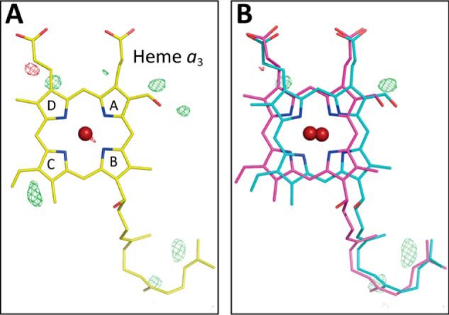
X-ray structure of heme a3 in the presence of 20 mm azide. A,Fo − Fc map determined assuming a single structure of heme a3. The residual densities are indicated with green mesh at +3.0 σ, and the converged structure is shown. B, Fo − Fc maps assuming the structures A and B of heme a3 (indicated by magenta and light blue sticks, respectively) in the 1:1 occupancy ratio. No significant residual density is detectable at the +3.0 σ level near the ring C.