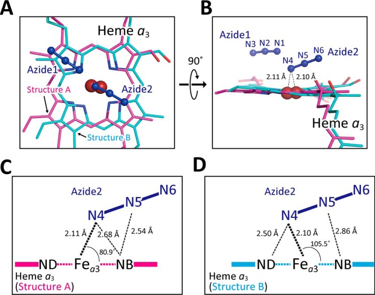
Figure 4.
Two modes of terminal azide bindings to Fea3 at 20 mm azide. A and B, atomic model of terminally-bound azide ions. Azide 1 and azide 2 in dark blue indicate azide ions bound to CuB and heme a3, respectively. Heme a3 planes giving structures A and B are drawn in magenta and light blue, respectively. C and D, geometry of bindings of azide 2 giving structures A and B, respectively, in the same color-code as in A and B.

