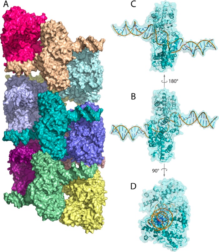
Figure 1.
A, surface rendering of oligomeric SgrAI–DNA (Protein Data Bank code 4C3G). Each SgrAI–DNA complex is colored a unique color. SgrAI is bound to two molecules of PC DNA forming a 40-bp duplex with nicks at the SgrAI cleavage sites. The oligomer has left-handed helical symmetry with approximately four SgrAI–DNA complexes per turn. B, one SgrAI–DNA complex in the same orientation as that in A, with cartoon rendering shown beneath the surface rendering (teal). Each subunit of the SgrAI dimer is shaded differently (light and dark teal). The DNA rendered in cartoon is colored yellow, green, and blue. C, the SgrAI–DNA complex shown in B, rotated 180° about the axis shown. D, the SgrAI–DNA complex shown in B, rotated 90° about the axis shown.

