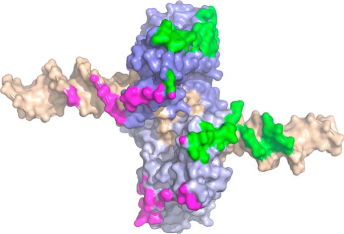
Figure 3.

Contact surface between SgrAI–DNA complexes of the run-on oligomer, mapped onto a single SgrAI–DNA complex. The two subunits of the SgrAI dimer are colored in different shades of blue, and the bound DNA (self-annealed PC DNA) is colored in wheat. Close contacts (within 4 Å) between SgrAI–DNA complexes in the ROO filament are limited to only the SgrAI–DNA complex just before (green) and just after (magenta) and occur on both the protein and DNA components of the SgrAI–DNA complex.
