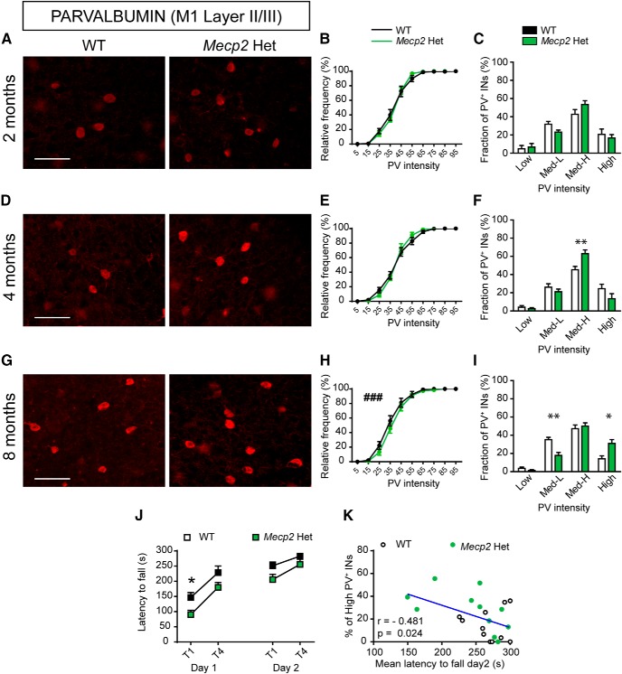
Figure 8.
Atypical high-PV expression in the M1 cortex of female Mecp2 Het mice correlates with motor impairments. Representative images showing PV expression in layer II/III of M1 cortex in both WT and Mecp2 Het mice at 2 (A), 4 (D), and 8 (G) months of age. Cumulative (B, E, H) and binned (C, F, I) frequency distribution of PV cells intensity in WT and Mecp2 Het M1 cortex at 2 (B, C), 4 (E, F), and 8 (H, I) months of age. J, Latency to fall (seconds) from an accelerating rotating rod in 8-mo-old Mecp2 Het mice and WT littermates. Graphs show data of first and last trials/d (T1–4), for two consecutive days (day 1–2). K, Correlation analysis between the mean latency to fall (seconds) from the rod on day 2 and the fraction of high PV+ INs in 8-mo-old Mecp2 Het and WT females. A–I: n = 6 WT and 6 Mecp2 Het mice; J, K: n = 11 WT and 11 Mecp2 Het mice. Mann–Whitney U test for B, E, H: ###p < 0.001; Student’s t test for C, F, I: *p < 0.05, **p < 0.01; two-way ANOVA and Bonferroni posthoc tests for J: *p < 0.05, **p < 0.01; Pearson’s r for E. Scale bar = 100 μm.

