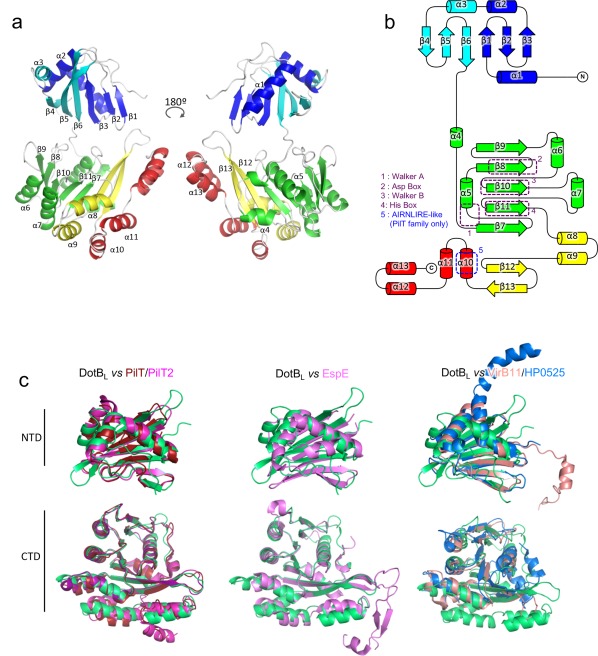
Figure 1.

DotBL crystal structure. (A) DotBL subunit structure. A single subunit in the α‐conformation is depicted in cartoon representation in two views rotated by 180°. Secondary structure elements are labeled and colored according to sub‐domains of the monomer. (B) Topology diagram of a DotBL subunit. β‐strands are represented by arrows, α‐helices by cylinders. The elements are colored as in a. Conserved regions are highlighted by dashed boxes. (C) Fold comparison of the NTD and CTD domains of DotBL with other secretion ATPases. Domains were aligned using the Cα of their conserved elements. From left to right: DotBL (green) aligned with PilT (red, pdb 2EWV) and PilT2 (magenta, pdb 5FL3), with EspE (pink, pdb 1P9R) and with B. suis VirB11 (light pink, pdb 2GZA) and HP0525 (blue, pdb 1NLY). The N‐terminal domain of DotBL aligns with that of Pilt2, PilT, HP0525, B. suis VirB11 and EspE with an rmsd in Cα atoms of 1.4, 2.0, 4.1, 4.9, and 1.4 Å, respectively while the C‐terminal domain aligns with that of Pilt2, PilT, HP0525, B. suis VirB11 and EspE with an rmsd in Cα atoms of 0.8, 0.8, 5.3, 3.3, and 1.8, respectively.
