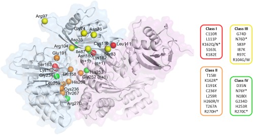
Figure 5.

Mapping of DotB mutants. Two adjacent subunits are depicted in cartoon and surface representations, with an α‐subunit in blue and the adjacent β‐subunit in purple. The locations of residues mutated in L. pneumophila DotB by Sexton et al. (2005) are identified by their Cα shown as spheres, color‐coded according to the phenotype classes listed on the right (see main text). The * indicates that the mutated residues are present in more than one class depending on the nature of the substitution.
