Figure 1.

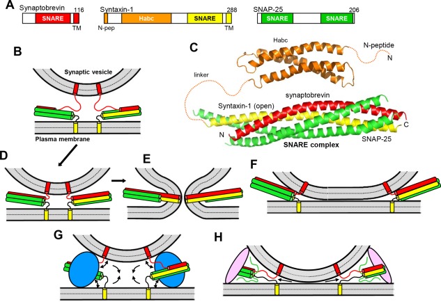
Models of SNARE‐dependent membrane fusion. (A) Domain structures of synaptobrevin, syntaxin‐1 and SNAP‐25. SNARE indicates SNARE motif and N‐pep indicates the N‐peptide of syntaxin‐1. Numbers on the right above the diagrams indicate the length of the protein. The same color coding for the SNAREs is used in all figures. (B) Diagram illustrating the topology of the neuronal SNAREs, with synaptobrevin anchored on a synaptic vesicle and syntaxin‐1 anchored on the plasma membrane, and showing how the SNAREs can form partially assembled trans‐SNARE complexes between the two membranes. In this model, the N‐terminal half of the four‐helix bundle is assembled and the C‐terminal half of the synaptobrevin SNARE motif is unstructured; the syntaxin‐1 and SNAP‐25 SNARE motifs are often assumed to be fully helical, as shown in the diagram, but their C‐termini may actually be unstructured.164 (C) Ribbon diagrams representing the three‐dimensional structures of the syntaxin‐1 H abc domain19 (PDB accession code 1BR0) and the neuronal SNARE complex12 (PDB accession code 1SFC). N and C indicate the N‐ and C‐terminus, respectively. Dashed curves represent flexible regions that were not present in the elucidated structures. (D–E) Together with panel (B), these diagrams illustrate the widespread model whereby the SNARE complex is initially formed at the N‐terminus (B), it zippers toward the C‐terminus, bringing the two membranes in close proximity (D), and causes membrane fusion as continuous helices are formed by the SNARE motifs and TM regions of syntaxin‐1 and synaptobrevin, as well as the short linkers between them.12, 27, 38 (E). For simplicity, only the SNARE motifs, TM regions and these short linkers are shown. (F) Diagram showing how SNARE complex assembly can lead to extended membrane‐membrane interfaces without fusion.32 (G) Diagram illustrating how a bulky protein(s) (blue) bound to the SNARE four‐helix bundle could play a key role in membrane fusion by pushing the membranes away from each other at the same time that assembly of the SNARE complex pulls the membranes together, which could cause a torque (see arrows) that helps to bend the membranes and initiate fusion.45, 46 (H) Diagram showing how membrane bridging by an elongated tethering factor (pink) could bring the vesicle and plasma membranes into proximity. In this arrangement, the SNARE complex would assemble in the periphery of the membrane‐membrane interface, perhaps bound to the bridging protein and/or another factor, and C‐terminal zippering of the SNARE complex could pull the membranes radially (see arrows), thus perturbing the packing of the lipids and catalyzing membrane fusion.48
