Figure 4.

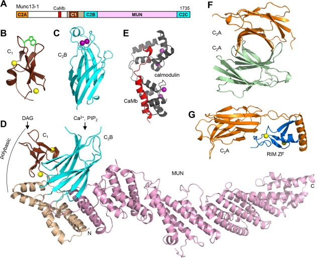
Structure of Munc13‐1. (A) Domain diagram of Munc13‐1. The calmodulin‐binding sequence is labeled CaMb. The number on the right above the diagram indicates the length of the protein. (B–G) Ribbon diagrams of the three‐dimensional structures of the C1 domain110 (B), the Ca2+‐bound C2B domain111 (C) and the C1C2BMUN fragment48 (D) of Munc13‐1, as well as calmodulin (purple) bound to the Munc13‐1 CaMb sequence (red)114 (E), the Munc13‐1 C2A domain homodimer116 (F) and the heterodimer of the Munc13‐1 C2A domain (orange) with the RIM2α ZF domain (blue)116 (G). The PDB accession codes are 1Y8F, 3KWU, 5UE8, 2KDU, 2CJT and 2CJS, respectively. Ca2+ ions are shown as purple spheres and zinc ions are shown as yellow spheres. In (D), the locations of the DAG/phorbol ester‐binding site in the C1 domain, the Ca2+/PIP2‐binding site in the C2B domain, and a polybasic region formed by the C1 domain, the C2B domain and the linker sequence between them are indicated.
