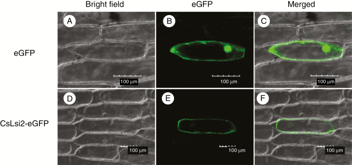
Fig. 3.
Subcellular localization of CsLsi2 in onion epidermal cells. The constructs carrying 35S::CsLsi1-eGFP or 35S::eGFP were transferred into onion epidermal cells. (A–C) Cellular localizations of CsLsi2; eGFP transformation in D–F was used as a positive control. A and D show bright-field images; B and E show eGFP fluorescence; and C and F show merged images. Scale bars = 100 μm.

