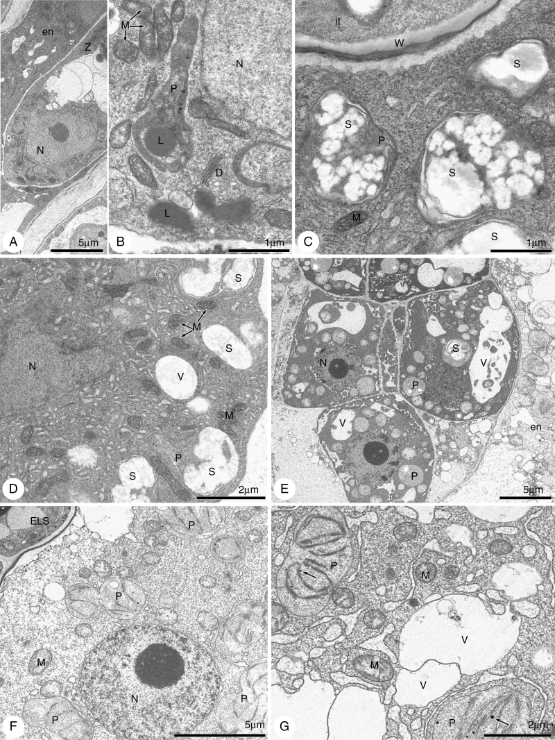
Fig. 11.
Electron micrographs of embryogenesis in line ES512. (A–D) A zygote and nuclear endosperm. Zygote cytoplasm (A, B) is dense and rich in lipids and long plastids without thylakoids, but accumulates lipids. The micropylar (C) and chalazal (D) cytoplasm of the nuclear endosperm is also dense and rich in organelles, especially in plastids accumulating huge popcorn-like starch grains. (E–G) Abnormal embryo and endosperm. (E) The irregularly positioned cells of the embryo have dark cytoplasm with non-typical starch-accumulating plastids and autophagic-like vacuoles. The micropylar endosperm (E, F) is degenerate and diluted, with disappearance of the organelles. The chalazal endosperm (G) is also diluted and fragmented but still contains mitochondria and spherical plastids with thylakoids. P, plastid; S, starch; V, vacuole; L, lipids; W, cell wall; N, nucleus; D, dictyosomes; en, endosperm; it, integumentary tapetum; z, zygote; M, mitochondria; ELS, embryo-like structure.

