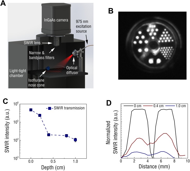
Figure 2.
A, Schematic of the small animal SWIR imaging system. B, PET chamber filled with REs and excited at 975 nm. Rod diameters counterclockwise from bottom are 4.8 mm, 4.0 mm, 3.2 mm, 2.4 mm, 1.6 mm, and 1.2 mm. C, Short-wave infrared signal from REs progressively diminished through increasing depth of phantom tissue, yet signal was still detected through 1.0 cm of tissue. D, Effects of increasing phantom depth on the SWIR resolution reveals point fluorescence could be distinguished with millimeter precision using the 4.8-mm-diameter rods as SWIR emission points. RE indicates rare-earth-doped nanoparticles; SWIR, short-wave infrared.

