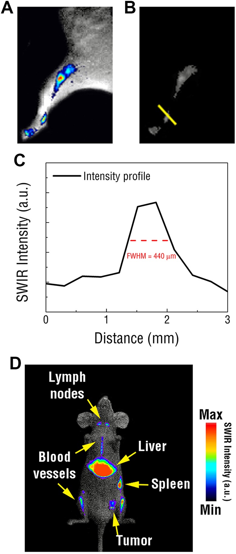
Figure 3.

A, Representative image showing tracking of the lymphatic vascular in the hind leg of a mouse using REs injected into the footpad. B, Raw SWIR image used to assess the resolution of SWIR signal tracked through the lymphatic vasculature. C, Reveals submillimeter, micron resolution. D, Representative SWIR image of PEGylated REs intravenously injected into a mouse exhibiting a 4T1 tumor reveals intense SWIR emissions emitted from the liver, spleen, and lymph nodes as well as certain vasculature and passive tumor accumulation. RE indicates rare-earth doped nanoparticles; SWIR, short-wave infrared; PEG, polyethylene glycol.
