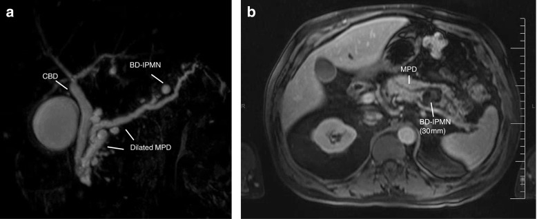
Fig. 1.

a MRCP—diluted pancreatic duct and Santorini with distal a diameter of less than 1 cm. Also, the image of multifocal small sidebranch IPMN. b MRI—ductus pancreaticus which is irregular at the level of the corpus and tail and is slightly dilated. Multiple cysteine deviations starting from the side duct. Largest cystic lesion located in the corpus with a staining solid component. Image matching a mixed-type IPMN with solid component as sign of a possible malignant degeneracy. PA—after pancreatic tail and spleen resection: the ductus pancreaticus and side branches show mixed-type IPMN, both gastric and pancreatobiliary type, with moderate dysplasia; there are extensive regressive changes with mucinous extravasation and fibrosis. No high-grade dysplasia, no malignancy
