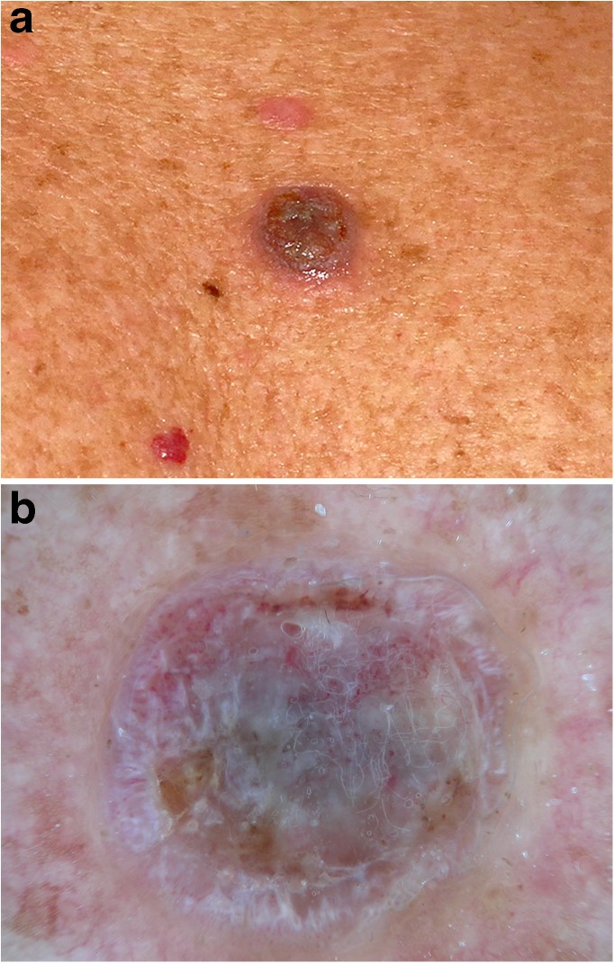
Fig. 6.
Hypopigmented nodule on the back of a 72-year-old man viewed with the unaided eye. a Dermatoscopic examination of lesion (b) shows ulceration with serum crusts and adherent fibers. Central gray color surrounded by white lines and coiled vessels allows the differential diagnosis of an amelanotic melanoma. Histopathologic diagnosis: nodular melanoma (Clark level, IV; Breslow depth, 2.4 mm; ulceration, stage T3b). Images courtesy of the Vienna Dermatologic Imaging Research Group, Department of Dermatology, Medical University of Vienna, Austria.

