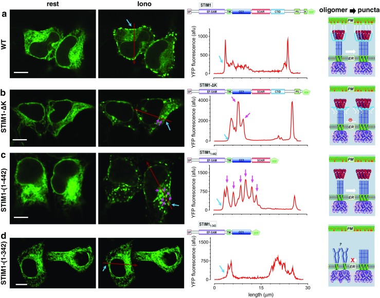
Fig. 2.

STIM1 protein without K-rich region could still form puncta in HEK Orai-KO cells. Different STIM1 constructs with YFP tagged at their C-terminus were transiently expressed in HEK Orai-KO cells and examined with confocal microscopy. Left, images of the middle plane of typical puncta-forming cells before (rest) and after store depletion (Iono: 5 min after 2.5 μM ionomycin treatments); scale bar, 10 μm; middle, profiles of YFP fluorescence along the red arrows (shown in images on the left) in store-depleted cells at two different focus planes. Red traces, in the middle plane of cells. Cell edges were indicated with blue arrows, and puncta formed outside of ER-PM junctions within cells were indicated with purple arrows. Right, diagrams showing proposed oligomerizing and clustering of STIM1 constructs deep within cells or at ER-PM junctions. a Full-length STIM1. STIM1 puncta are mostly localized on the peripheral of the cells. b STIM1-ΔK. In all the cells expressing STIM1-ΔK we examined, about 5% of them could form sparse puncta after store depletion. Without the help of PM-anchoring poly-K region, some STIM1 puncta are located within the interior of cells (indicated by purple arrows). c STIM1-(1-442). Without the entire region C-terminal to SOAR/CAD, massive STIM1 puncta were formed all over the cell. d STIM1-(1-342). After the deletion of SOAR/CAD region, STIM1 lost its ability to form puncta after store depletion. The densities of intracellular clusters that were formed outside of ER-PM junctions were 0, 1.6 ± 0.4, 13.2 ± 3.2, and 0% for wt, STIM1-ΔK, STIM1-(1-442) and STIM1-(1-342), respectively (n = 3, at least six puncta-forming cells were examined each time)
