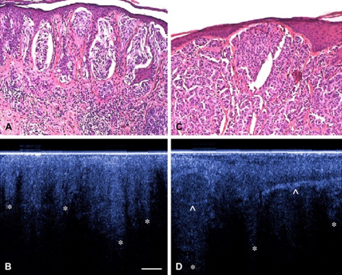
Fig. 2.
Example of corresponding OCT and histology images of two melanomas Upper panel (a, c): Hematoxylin and eosin (H&E) images of a superficial spreading melanoma. Lower panel (b, d): OCT images of distorted skin architecture, including large vertically arranged icicle-shaped structures (*). Prominent hyperreflective structures are corresponding to dense collagen cords of encapsulated tumor lobules.
Reprinted by permission from Elsevier: Journal of the American Academy of Dermatology (Gambichler et al. 2007). © 2007

