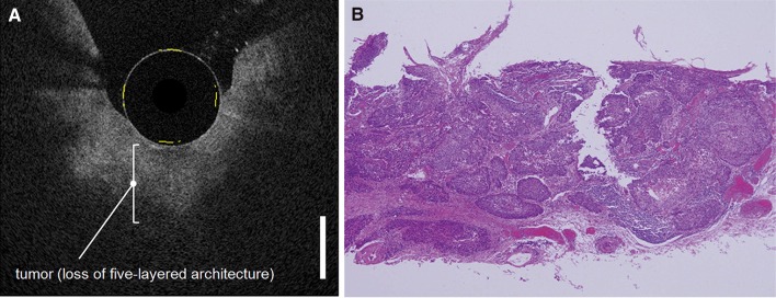
Fig. 5.
Example of endoscopic OCT of an esophageal squamous cell carcinoma. Corresponding OCT (a) and histology (b) image of tumor invasion in the submucosal layer, resulting in a loss of the five-layered architecture (Bar = 1000 µm).
Reprinted by permission from Elsevier: Gastrointestinal Endoscopy (Hatta et al. 2010). © 2010

