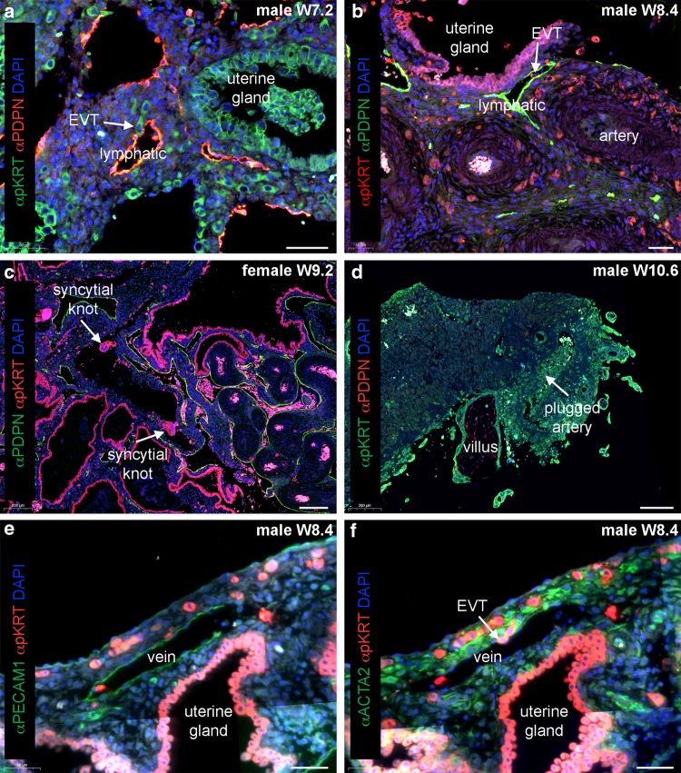
Fig. 2.
Endovenous and endolymphatic trophoblast invasion in normal pregnancies. Sections from first trimester decidua basalis (gestational age 7.2–10.6 weeks). a Immuno-double staining for pan cytokeratin (pKRT, green) and podoplanin (PDPN, red) with nuclear counterstain (DAPI, blue). b Immuno-double staining for pKRT (red) and PDPN (green) with DAPI (blue). White arrows in a, b depict endolymphatic trophoblasts invading lymphatic vessels. c Immuno-double staining for pKRT (red) and PDPN (green) with DAPI (blue). White arrows depict syncytial knots within a uterine vein. d Immuno-double staining for pKRT (green) and PDPN (red) with DAPI (blue). White arrow depicts a plugged artery. e, f Immuno-double staining for e pKRT (red) and PECAM1 (green) with DAPI (blue) and an adjacent section f with immuno-double staining for ACTA2 (green) and pKRT (red) with DAPI (blue). White arrow depicts endovenous trophoblasts invading a vein. EVT, extravillous trophoblast. Scale bars represent 50 µm in a, b, e, f and 200 µm in c, d

