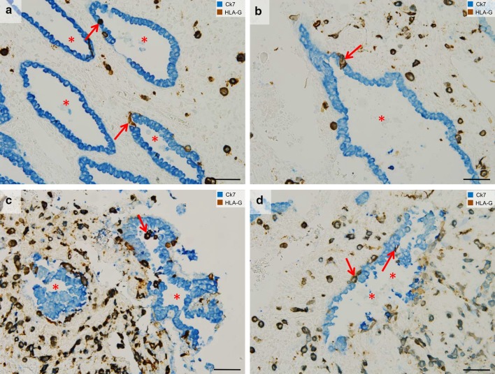
Fig. 3.
Endoglandular trophoblast invasion. Sections from the first trimester decidua basalis (gestational age 7–8 weeks). a–d Immuno-double staining for cytokeratin 7 (blue) and HLA-G (appears brown/dark violet) without nuclear counterstain. a, b Endoglandular trophoblasts (arrows) replace the epithelium of uterine glands (lumen of gland: asterisk). c Endoglandular trophoblasts are also situated in the glandular lumen. d Epithelium of invaded glands often appears disintegrated. Endoglandular trophoblasts, arrows; lumen of gland, asterisk. Scale bars represent 50 µm

