Fig. 4.
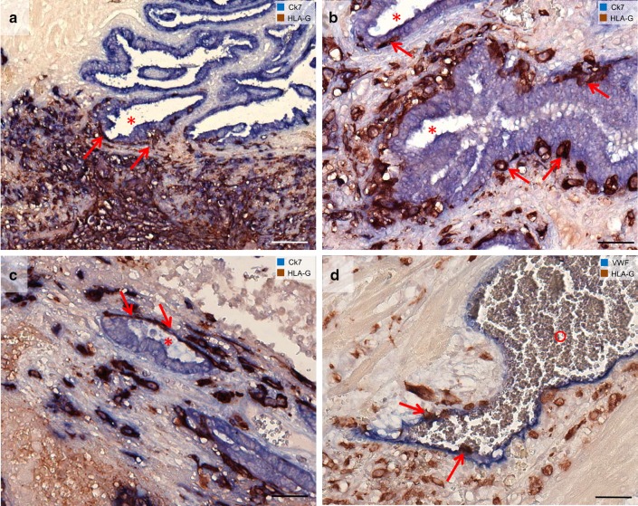
Invasion of extravillous trophoblasts in epithelial structures and vessels in tubal pregnancies. Sections from tubal pregnancies (a–c) are immuno-double stained for cytokeratin 7 (blue) and HLA-G (appears brown/dark violet) without nuclear counterstain. a Overview. Extravillous trophoblasts (arrows) invade the lamina propria of the mucosal folds (asterisk). Trophoblasts can be found beneath the epithelium. b, c The epithelium of the mucosal folds is penetrated by extravillous trophoblasts (arrows) from the basal side. d Extravillous trophoblasts are visualized with immuno-double staining for vWF (blue) and HLA-G (brown). Single endovascular trophoblasts (d, arrows) are situated in the lumen of the vessel (circle). Scale bars represent 100 µm in a and 50 µm in b, c, d

