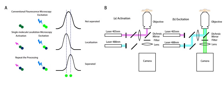
Figure 1.
Schematics of the working principle and the setup of the stochastic optical reconstruction microscopy (STORM). (A) The top row shows the two fluorophores (located along the two dashed lines) activated and excited simultaneously by conventional microscopy. Due to the diffraction limit, the two fluorophores cannot be separated, resulting in a blurring image. The middle and bottom rows show the principle of STORM for the localization of a single molecule to a nanometer accuracy. The fluorophores are activated and excited not simultaneously, but sequentially to be localized and separated with each other. This technique can overcome the diffraction barrier for the conventional fluorescence microscopy. (B) A representative optical set up of STORM for the fluorophore Auto 488. (a) The optical path in activation process. The 405nm wavelength laser activates the photo-switchable fluorophore Auto 488, enabling the excitation. (b) The optical path in excitation and imaging processes. The 488nm laser excites the activated “on” fluorophore and its emission is recorded by a camera.

