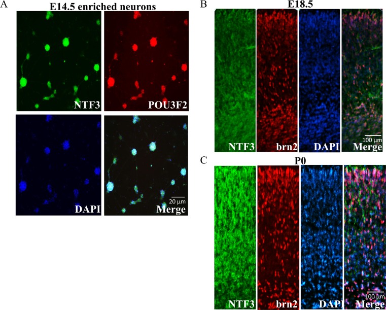
Fig. 6.
Colocalization of POU3F2 and NTF3 proteins in the developing mouse neurons. a Primary mouse neurons (EN) were prepared from E14.5 embryos, cultured for 7 days, and subjected to double fluorescence immunostaining using anti-NTF3 (green) and anti-POU3F2 (red) antibodies and FITC. Immunostaining was also performed on cortical sections of mouse brain prepared from E18.5 embryos (b) and postnatal day (P) 0 mice (c). Merged images of NTF3 and POU3F2 showed extensive overlap of expression in primary enriched cultured neurons and in the E18.5 and P0 cortex, mainly at the upper and deeper layers

