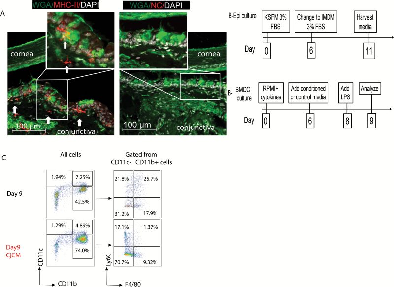
Fig. 1.
BMDC culture phenotype and suppressive effects of CjCM. (A) Representative laser confocal images of conjunctival section co-stained with anti-MHCII (red), WGA lectin (green) that binds to goblet cell glycoproteins and DAPI for nuclear DNA (white). Arrows point to MHCII+ dendritic-shaped APCs below the epithelium and in the superficial stroma. Inset in left image of MHCII+ DC adjacent to WGA+ goblet cell in basal conjunctival epithelium. NC = negative control with second antibody only. (B) Experimental design for preparation of conjunctival (goblet cell) epithelial conditioned media (Epi culture, upper) and BMDC culture (lower) treatment groups. (C) Representative density plots of BMDCs cultured in control IMDM media (top) or in conjunctival (goblet cell) epithelial conditioned media (CjCM, bottom) from days 6 to 9. The plots on the right were gated from the CD11c−CD11b+ cells. Day 9 = control BMDCs; Day 9 CjCM = BMDCs treated with conjunctival (goblet cell) epithelial conditioned media; Ly6C = monocyte marker; F4/80 = macrophage marker.

