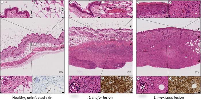
FIG 7.
Comparison of mouse skin morphology and macrophage density in healthy, uninfected skin (left), L. major CL lesion (20 days postinfection, middle), and L. mexicana CL lesion (45 days postinfection, right). The central picture in each panel (H&E stain) shows the structural layers of the skin, epidermis (E), dermis (D) and hypodermis (H), with the underlying muscle (M) at ×4 magnification (bar = 100 μm). The insets (1 to 4) highlight details of the central picture (×80 magnification, bar = 10 μm). ①, epidermis; ②, dermal capillaries; ③, Leishmania amastigotes within parasitophorous vacuoles; ④, anti-Iba-1 stain (macrophage marker) of tissue shown in inset ③. In both the L. major and L. mexicana CL lesions, intense inflammatory foci (I) are present in the skin, causing severe disruption of the D and H architecture. Compared to healthy uninfected skin, CL lesions also showed (i) epidermal hyperplasia and acanthosis for L. mexicana but not for L. major (①), (ii) dilated blood vessels, a factor contributing to capillary leakiness (②), and (iii) a large amount of inflammatory cells (③), many of which are macrophages (④).

