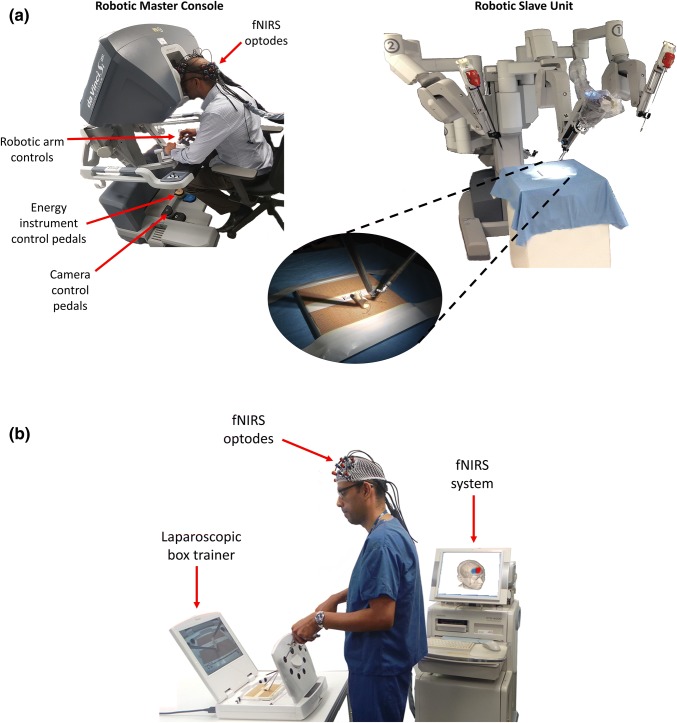
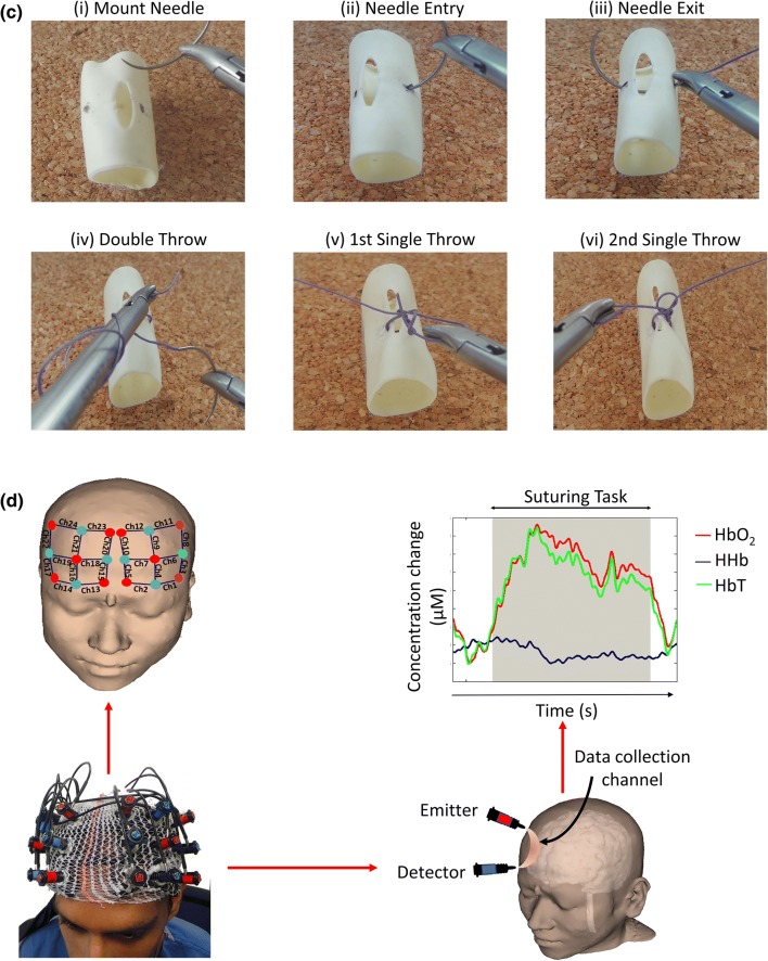
Figure 1.
(a) The da Vinci® Si system (Intuitive Surgical Inc, Sunnyvale, CA) consists of a master console system with which the surgeon controls a slave unit comprising robotic arms that move around fixed pivot points and carry out the surgeon’s commands; (b) A bench-top box trainer (iSim2, iSurgicals, UK) used for the laparoscopic suturing task; (c) Key steps of the suturing task in which a reef knot is created: (i) mounting the needle onto the needle holder, (ii) inserting the needle into the Penrose drain as close to pre-marked target points as possible, (iii) exiting the needle out of the drain as close to pre-marked target points as possible, (iv) double throw of suture thread, (v) first single throw, and (vi) second single throw; (d) Prefrontal activation during the task was assessed using functional near-infrared spectroscopy (fNIRS), a non-invasive neuroimaging technique, which measures differences between emitted and detected near infrared light to estimate the local concentration changes of oxygenated haemoglobin (HbO2) and deoxygenated haemoglobin (HHb), as a surrogate of brain activation. The 3D head reconstruction demonstrates the 3 × 3 arrays of optodes located over the left and right PFC, along with the positions of 24 corresponding channels (‘Ch’) that measure haemodynamic responses in an area of cortex located between an emitter (red) and detector (blue). The typical cortical haemodynamic response in channels exhibiting activation comprises an increase in HbO2, a smaller decrease in HHb, and an increase in total haemoglobin (HbT = HbO2 + HHb).


