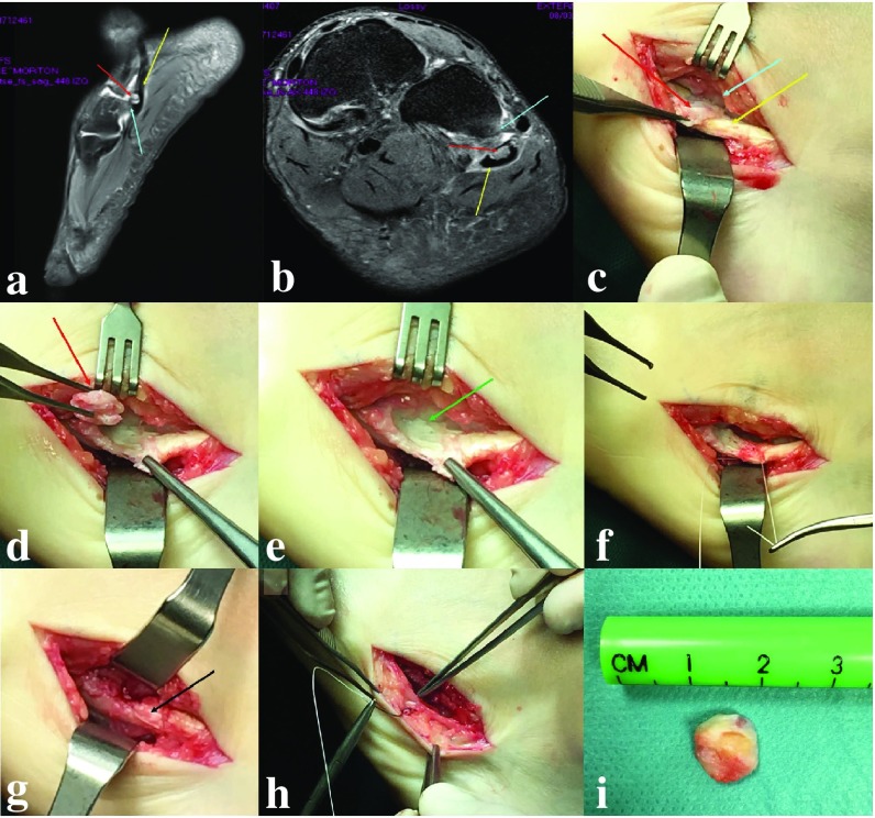
Fig. 6.
Case of symptomatic Os Peroneum. Peroneum causing impingement with the cuboid. Os Peroneum (OP—red arrows); Peroneus Longus (PL—yellow arrows) on lateral (a) and axial (b) MRI views. In the conflict area of the OP with the cuboid it is visible some bone edema in T2 MRI sequences (light blue arrows). c PeroperaCve image with visibility of the OP (red arrow), PL (yellow arrow) and impingement area with the cuboid (light blue arrow). The OP is detached from the PL keeping the integrity of the PL. The peroneal Cssue is flaIened (green arrow) in the zone where the OP was removed (e). f Reinforcement sutures of the PL are performed with tubularizaCon of the flaIened area (g—black arrow). h Be aware of the close connecCon with the sural nerve (pointed by surgical tweezers) during all the procedure and confirm its integrity in the end before closure of the wound. i OP after removal in one piece

