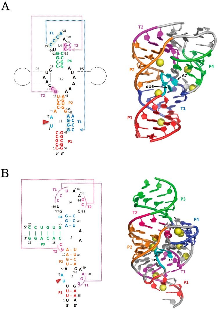
Figure 1.
Secondary and tertiary structures of two representatives of twister ribozymes. (A) The structure of the twister ribozyme from O. sativa [30]. Additional stem-loop segments, P3 and/or P5, can be generated, as shown in black dotted lines; (B) The structure of the env22 twister ribozyme [29]. In (A) and (B), red arrowhead indicates the U-A cleavage site. On the secondary structure, highly conserved nucleotides (>97%) are marked by asterisks. Stems (P1-P4) and pseudoknots (T1 and T2) are colour-coded in the tertiary structure. In particular, two nucleotides at the cleavage site and bound magnesium ions are coloured in cyan and yellow, respectively. Protein Data Bank (PDB) accession codes for (A) and (B) are 4OJI and 4RGE, respectively.

