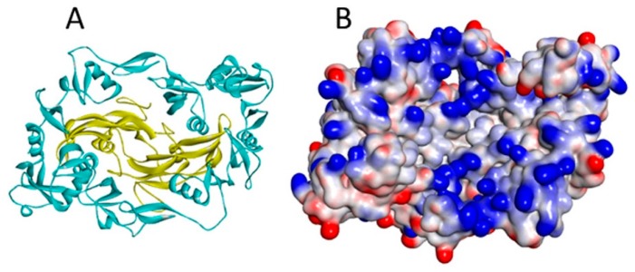
Figure 5.
(A) The myostatin (yellow ribbon)/follistatin (turquoise ribbon) complex (3HH2.pdb); and (B) the same complex shown as a surface coloured according to interpolated charge (positive is blue, negative is red). Though myostatin does not have a heparin binding site, basic residues on its surface are located close to the follistatin binding site in the complex, increasing total affinity for heparin [40].

