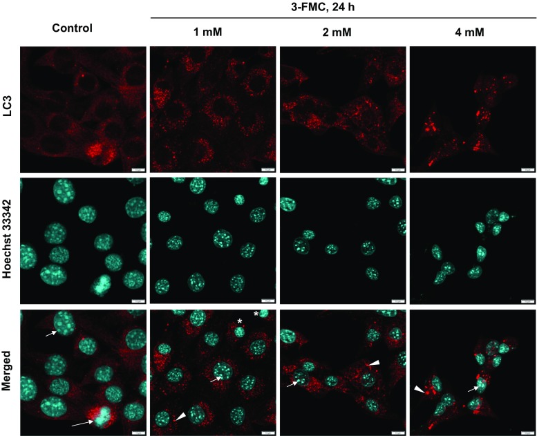
Fig. 3.
Immunofluorescent analysis. Confocal micrographs of HT22 cells treated with 1, 2, and 4 mM 3-FMC for 24 h. Cells were incubated with primary anti-LC3 antibodies. Following incubation with Cy3-conjugated secondary antibodies and Hoechst 33342, cells were examined by confocal microscopy as described in Materials and Methods. Data are representative of three independent experiments. Bars 10 μm, control—untreated cells, arrowheads—autophagic vacuoles, short arrows—nucleoli, long arrow—a cell undergoing mitosis, asterisks—newly formed cells after cell division

