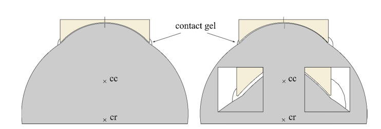
Fig. 3.
True to scale sections of eyeball, cornea and concave TomoCap with a gel interface of 50 µm thickness. The cornea’s center of curvature is denoted with cc and the eye’s center of rotation with cr. Vertical lines on the cornea apex and center of the concave surface visualize the rotation of the eye. Left: no rotation; Right: Clockwise rotation around cr of 0.66 ° corresponding to a lateral corneal shift of 150 µm. Insets show the distance between cornea and TomoCap and schematically the amount of surrounding gel.

