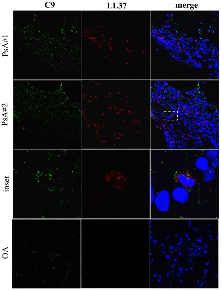
Figure 10.
C9 staining in synovial tissues of PsA. Confocal microscopy images of synovial tissues of two PsA and one OA patients stained for LL37 (red) and C9 (green) (original magnification, 63x). The dotted white line in PsA#2 indicates the inset of the corresponding picture of the lower panel. Data are representative of 7 PsA patients and 4 OA subjects.

