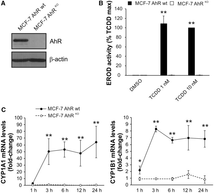
Figure 1.
Lack of inducible CYP1A1 and CYP1B1 expression/activity in MCF-7 AhRKO cells. A, MCF-7 AhRKO cells were routinely monitored for the lack of AhR expression throughout the study. A representative result of Western blotting detection of AhR protein in MCF-7 AhR wt and MCF-7 AhRKO cells is shown. B, TCDD failed to induce detectable 7-ethoxyresorufin-O-deethylase (EROD) activity in MCF-7 AhRKO cells. Cells were incubated with DMSO (0.1%; negative control) or indicated concentrations of TCDD for 24 h and EROD assay was then performed as described in Materials and Methods. The results represent means ± SD of three independent experiments. Symbol “**” denotes significant difference (p < 0.01) between MCF-7 AhR wt and MCF-7 AhRKO cells for the respective treatment group. C, BaP-induced CYP1A1 and CYP1B1 mRNA in MCF-7 AhR wt cells but not in MCF-7 AhRKO cells. Cells were exposed to BaP (10 μM) for indicated time, lysed and total RNA was isolated. CYP1A1/1B1 mRNA levels were determined using qRT-PCR as described in Materials and Methods. The results represent means ± SD of three independent experiments. Symbol “*” denotes significant difference (p < .05) between MCF-7 AhR wt and MCF-7 AhRKO cells at the respective time-point. Symbol “**” denotes significant difference (p < .01) between MCF-7 AhR wt and MCF-7 AhRKO cells at the respective time point.

