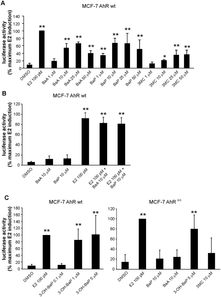
Figure 4.
Effects of PAHs on induction of estrogen-dependent luciferase reporter gene in MCF-7 AhR wt and MCF-7 AhRKO cells. A, MCF-7 AhR wt cells were grown in experimental medium for 24 h and then transiently transfected with 3X ERE TATA luc reporter construct and pRL-TK vector, encoding Renilla luciferase (transfection efficiency control). After 24 h of transfection, cells were exposed to DMSO (0.1%; negative control), E2 (100 pM; positive control), or indicated concentrations of PAHs for 24 h. Following the incubation, cells were collected, lysed and firefly/Renilla luciferase activities were determined with a luminometer. The results were expressed relative to maximum luciferase activity induced by reference compound (E2). B, MCF-7 AhR wt cells were grown in experimental medium for 24 h and then transfected with 3X ERE TATA luc reporter construct and pRL-TK vector as above. Aftere 24 h of transfection, cells were exposed to DMSO (0.1%; negative control), E2 (100 pM; positive control), BaA and BaP, or combinations of E2 and the respective PAH, for 6 h. Following the incubation, cells were collected, lysed, and firefly/Renilla luciferase activities were determined with a luminometer. C, MCF-7 AhR wt cells and MCF-7 AhRKO cells were grown in experimental medium for 24 h and then transfected with 3X ERE TATA luc reporter construct and pRL-TK vector as above. After 24 h of transfection, MCF-7 AhR wt cells (left panel) were exposed to DMSO (0.1%; negative control), E2 (100 pM; positive control), or indicated concentrations of 3-OH-BaP for 24 h. MCF-7 AhRKO cells (right panel) were exposed to DMSO, E2 or indicated concentrations of BaA, BaP, 3-OH-BaP, and 3MC for 24 h. Following the incubation, cells were collected, lysed, and firefly/Renilla luciferase activities were determined with a luminometer. All results were expressed relative to maximum luciferase activity induced by reference compound (E2). The results shown here represent means ± SD of three independent experiments. Symbol “*” denotes significant difference (p < .05), as compared with DMSO-treated cells. Symbol “**” denotes significant difference (p < .01), as compared with DMSO-treated cells.

